
INTRODUCTION
Prosthetic valve endocarditis (PVE) caused by nontuberculous mycobacteria (NTM) is rare, diagnostically challenging, and associated with substantial morbidity and mortality.1,2 Among rapidly growing mycobacteria, PVE has been more frequently attributed to organisms such as Mycobacterium chelonae, Mycobacterium fortuitum, and Mycobacterium abscessus, whereas organisms within the Mycobacterium smegmatis group have only rarely been implicated.2,3 Mycobacterium smegmatis sensu stricto, a member of the Mycobacterium smegmatis group, is itself an exceptionally uncommon cause of invasive human disease, and the limited historical endocarditis literature largely predates modern species-level classification within the Mycobacterium smegmatis group.4–6 Mycobacterium goodii, also a member of the Mycobacterium smegmatis group and first described in 1999, has been associated primarily with post-traumatic wound infections, orthopedic hardware infections, and osteomyelitis; cardiovascular involvement remains exceedingly rare.6–8
Published reports of prosthetic valve endocarditis involving organisms within the Mycobacterium smegmatis group and closely related rapidly growing mycobacteria are limited (Table 1).5,7–9 Two cases were attributed to Mycobacterium goodii. Both involved mitral prosthetic material and neurologic septic embolic complications and were managed with surgical replacement and prolonged combination antimicrobial therapy.7,8 A third case involved Mycobacterium wolinskyi in a patient with multiple cardiac prostheses; routine identification was nondiagnostic, sequencing was required for species identification, and the infection was ultimately controlled with medical therapy alone.9 An older postoperative case reported as Mycobacterium smegmatis predates modern taxonomic subdivision of the group.5,6 The present case is therefore best classified as another prosthetic valve endocarditis case involving the Mycobacterium smegmatis group, without a specific ordinal designation. The blood isolates were reported as Mycobacterium smegmatis, whereas the explanted valve isolate was reported as Mycobacterium goodii. Because both reported identifications fall within the Mycobacterium smegmatis group and definitive species-level confirmation was unavailable, the infection is reported at the group level.6 Unlike the previously reported Mycobacterium goodii cases, the present case involved a bioprosthetic aortic valve with additional left atrial appendage occlusion hardware and remained echocardiographically nondiagnostic despite persistent mycobacterial bacteremia.7,8
Nontuberculous mycobacterial prosthetic valve endocarditis may present subacutely with nonspecific symptoms and persistent or intermittent bacteremia.10 Standard echocardiographic evaluation may repeatedly be nondiagnostic, particularly when infection involves prosthetic material or periannular structures.11,12 Advanced imaging is therefore often used when clinical suspicion for PVE remains despite negative or inconclusive echocardiography.11,12 Current diagnostic frameworks support this approach: the 2023 European Society of Cardiology (ESC) guidelines recommend fluorine-18 fluorodeoxyglucose positron emission tomography/computed tomography (18F-FDG PET/CT) in patients with suspected PVE when echocardiography is inconclusive, and the 2023 Duke–International Society for Cardiovascular Infectious Diseases (ISCVID) criteria incorporate abnormal 18F-FDG PET/CT metabolic activity involving a prosthetic valve, intracardiac device leads, or other prosthetic material as a major imaging criterion when PET/CT is performed at least 3 months after implantation of the prosthetic material.11,13 In this context, PET/CT can help localize metabolically active prosthetic infection, identify extracardiac foci, and guide decisions regarding surgical source control.11,12
In parallel with evolving diagnostic strategies, therapeutic options for rapidly growing NTM infections remain limited by antimicrobial resistance patterns and medication intolerance, particularly when prosthetic material is involved.10,14 Clofazimine, historically used in leprosy, has been used as an adjunct in multidrug regimens for NTM infections based on in vitro activity and limited clinical experience; however, evidence supporting its use in NTM endocarditis remains very limited, and its use in this setting is off-label.15–17 We report a case of Mycobacterium smegmatis group prosthetic aortic valve endocarditis with dual prosthetic involvement, in which serial transesophageal echocardiography was nondiagnostic and PET/CT contributed important diagnostic information that supported surgical source control.
An 82-year-old man with severe aortic stenosis underwent bioprosthetic aortic valve replacement with a 25-mm Magna Ease valve and left atrial appendage occlusion using an AtriClip device in April 2021. His relevant medical history included hypertension, hyperlipidemia, hypothyroidism, stage 2 chronic kidney disease, hiatal hernia, iron-deficiency anemia, prior squamous cell carcinoma, and remote tobacco use. He had no documented history of diabetes mellitus, immunosuppressive therapy, or prior mycobacterial bacteremia. Baseline outpatient medications included amlodipine 10 mg once daily and levothyroxine 25 µg once daily, along with vitamin and iron supplementation. Before the onset of illness, the patient lived at home with his wife, was ambulatory without assistance, and was independent in activities of daily living. Following surgery, his cardiovascular status improved, but he did not return to his previous functional baseline. He subsequently developed recurrent fevers beginning in late July or early August 2021.
At that stage, the differential diagnosis remained broad and included occult postoperative infection, noncardiac infectious sources, noninfectious causes of fever, and prosthetic valve endocarditis. A timeline of the major diagnostic and therapeutic events is presented in Table 2.
In December 2021, blood cultures were intermittently positive for Mycobacterium smegmatis, with positive cultures interspersed with negative routine and acid-fast bacillus (AFB) blood cultures through April 2023 (Table 3).
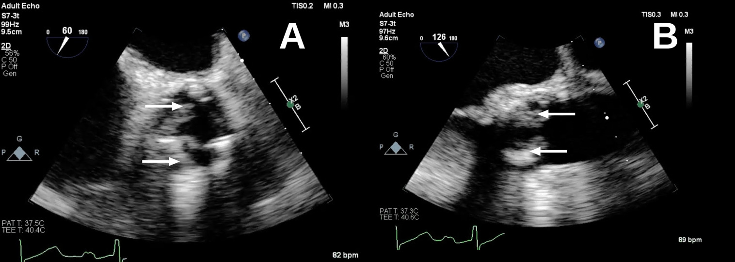
Concurrent transesophageal echocardiography (TEE) performed in December 2021 demonstrated abnormally thickened prosthetic valve leaflets but no definitive vegetations or periannular complications (Figure 1).
_view_obtained_at_a_multiplane_angle.png)
Taken together, these findings did not establish a definitive diagnosis of PVE. Rapidly growing NTM are uncommon causes of endovascular infection.10,14 The bacteremia was intermittent, and TEE did not demonstrate vegetations or periannular complications. At that time, contamination, transient bacteremia, and noncardiac sources of fever remained plausible considerations. Accordingly, immediate reoperation was not pursued. Instead, the patient was managed with antimicrobial therapy and close clinical and microbiological surveillance. Initial therapy consisted of a 6-week course of intravenous meropenem, oral doxycycline, and oral ethambutol, followed by an outpatient oral regimen, as summarized in Table 2.
During this period, brain magnetic resonance imaging (MRI), obtained in December 2021 for evaluation of headache, demonstrated a tiny acute ischemic infarct in the inferior right cerebellum without hemorrhage. At that time, the lesion was considered an incidental finding and was not attributed to septic embolization because PVE had not yet been established.
Antimicrobial therapy was briefly discontinued around April 2022, but symptoms recurred within several weeks, prompting resumption of doxycycline, clarithromycin, and ethambutol. Doxycycline was subsequently replaced with trimethoprim-sulfamethoxazole (TMP-SMX), although the exact durations of these outpatient regimens were not consistently documented. Despite ongoing treatment, blood cultures remained intermittently positive.
A repeat TEE in September 2022 demonstrated a well-seated, normally functioning bioprosthetic aortic valve without vegetations, preserved biventricular systolic function, and no intracardiac thrombus. Given the absence of definitive echocardiographic evidence of PVE, the patient continued to be managed with antimicrobial therapy, surveillance blood cultures, and close follow-up rather than undergoing immediate reoperation. However, recurrent fevers and intermittent mycobacterial bacteremia (Table 3) persisted despite multiple antimicrobial regimens, making contamination or transient bacteremia less likely and increasing concern for occult prosthetic valve or device-related infection not adequately visualized by echocardiography.
Antimicrobial susceptibility testing of the blood isolate, performed in February 2023, showed low minimum inhibitory concentrations (MICs) for several oral agents, including linezolid, moxifloxacin, minocycline, and clofazimine (Table 4).
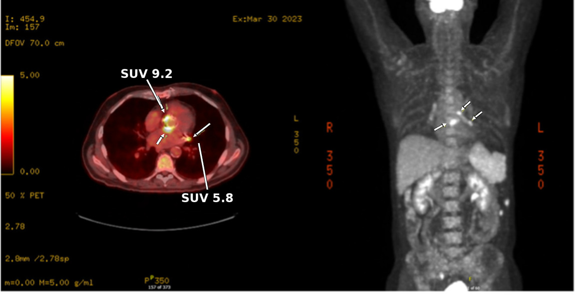
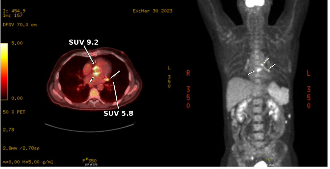
Persistent bacteremia and recurrent fevers prompted advanced imaging with PET/CT in March 2023, which demonstrated intense uptake surrounding the aortic valve prosthesis (maximum standardized uptake value [SUV] of 9.2) and focal uptake at both ends of the AtriClip device (SUV of 5.8), raising concern for infectious or inflammatory involvement of prosthetic material (Figure 2).
Because recurrent fevers and mycobacterial bacteremia persisted despite the preceding multidrug regimen, the infectious diseases team changed therapy in April 2023 to linezolid 600 mg once daily, moxifloxacin 400 mg once daily, and minocycline 100 mg twice daily. This regimen was consistent with the February 2023 susceptibility profile, which demonstrated low MICs for linezolid (<1 µg/mL), moxifloxacin (<0.5 µg/mL), and minocycline (<0.5 µg/mL).
Given persistent mycobacterial bacteremia and PET/CT findings localizing suspected involvement of the aortic valve prosthesis and left atrial appendage occlusion hardware, the anticipated benefits of repeat valve surgery were judged to outweigh the considerable operative risks, including those associated with the patient’s advanced age.
On May 9, 2023, the patient underwent redo sternotomy with explantation of the infected bioprosthetic aortic valve, aggressive aortic root debridement and circumferential reconstruction using a bovine pericardial patch, and implantation of a 25-mm bioprosthetic valve. The AtriClip device was removed, and the left atrial appendage was oversewn.
Intraoperative findings demonstrated a grossly infected prosthetic valve with thickened and discolored leaflets and numerous small vegetations. A circumferential abscess cavity was present within the aortic root, with destruction of the entire aortic annulus, the aorto-mitral continuity, and most of the left ventricular outflow tract, necessitating aggressive debridement and circumferential bovine pericardial reconstruction. Operative specimens were sent for routine bacterial, anaerobic, fungal, and acid-fast bacillus (AFB) cultures. Routine bacterial, anaerobic, and fungal cultures showed no growth. AFB culture of the explanted valve grew a rapidly growing mycobacterium. At that time, the valve isolate was reported as Mycobacterium goodii based on matrix-assisted laser desorption/ionization time-of-flight mass spectrometry (MALDI-TOF). Because Mycobacterium goodii is a member of the Mycobacterium smegmatis group,6 this identification was consistent at the group level with the patient’s earlier blood isolates reported as Mycobacterium smegmatis. Surgical pathology of soft tissue from the explanted AtriClip device demonstrated a foreign body giant cell reaction.
The postoperative course was complicated by complete atrioventricular block with pacing dependence, requiring implantation of a Micra AV leadless permanent pacemaker in May 2023. The postoperative regimen required modification because of lactic acidosis, thrombocytopenia, and hepatotoxicity. Oral clofazimine (100 mg once daily) was incorporated into the prolonged multidrug therapy because susceptibility testing demonstrated a low MIC and other standard oral options were limited by toxicity or poor tolerability. The patient was discharged in June 2023 on a clofazimine-containing multidrug antimicrobial regimen, which was continued for approximately five months after hospital discharge.
Follow-up routine and acid-fast bacillus (AFB) blood cultures, obtained from May 10 through September 13, 2023, showed no growth (Table 5). No subsequent microbiologic evidence of recurrence was documented. Fever resolved following surgical source control and completion of antimicrobial therapy.
A transthoracic echocardiogram showed eccentric mitral regurgitation that was difficult to quantify. Cardiac magnetic resonance imaging performed in February 2024 demonstrated moderate mitral regurgitation, a preserved left ventricular ejection fraction of 57%, and no evidence of prosthetic aortic valve dysfunction.
At the most recent follow-up in May 2026, approximately three years after redo aortic valve surgery, the patient remained asymptomatic with no clinical evidence of recurrent infection. Since discharge, he has experienced no infection-related rehospitalizations, has returned to his baseline functional status, and has remained independent in activities of daily living. He continued routine follow-up with cardiology and primary care.
PATIENT PERSPECTIVE
From the patient’s perspective, the prolonged diagnostic course was characterized primarily by fatigue and reduced physical reserve during recurrent febrile episodes. Following definitive treatment, he reported substantial improvement in quality of life and a return to baseline functional status.
DISCUSSION
This case highlights the diagnostic challenges of PVE caused by rapidly growing NTM in the setting of persistent or relapsing bacteremia and repeatedly nondiagnostic echocardiography. Although TEE remains central to the evaluation of suspected PVE, its sensitivity may be limited when infection involves prosthetic material, periannular tissue, or early abscess formation without a discrete vegetation.11,12 In the present case, serial TEE demonstrated only subtle prosthetic leaflet thickening without definitive evidence of infective endocarditis, whereas operative findings revealed extensive prosthetic valve infection and a circumferential aortic root abscess. This discordance represents a key feature of this case and reinforces that negative or equivocal echocardiographic findings should not be used to exclude prosthetic valve infection when persistent mycobacterial bacteremia is present.
The present case shared several features with the Mycobacterium goodii prosthetic valve endocarditis cases reported by Jönsson et al. and Parikh and Grant: a subacute presentation, involvement of prosthetic cardiac material, possible neurologic embolization, and the need for prolonged multidrug antimicrobial therapy and surgical source control.7,8 Given the limited literature on prosthetic valve endocarditis involving the Mycobacterium smegmatis group and closely related rapidly growing NTM, this case adds to the literature by illustrating an atypical presentation characterized by equivocal TEE findings and intermittent bacteremia.5,7–9 In retrospect, the cerebellar infarct may have represented an embolic phenomenon associated with the subsequently confirmed prosthetic valve and device infection, although a definitive embolic source was not established in the present case.
In contrast, both prior cases involved mitral prosthetic material, whereas the present case involved a bioprosthetic aortic valve with additional left atrial appendage occlusion hardware. Unlike the previously reported Mycobacterium goodii prosthetic valve cases, in which TEE demonstrated vegetations or other definitive prosthetic involvement, serial TEE in the present case remained nondiagnostic despite persistent mycobacterial bacteremia.7,8 PET/CT localized suspected prosthetic infection, which was ultimately confirmed surgically.
In this case, PET/CT contributed important diagnostic information by localizing metabolically active abnormalities involving both the aortic valve prosthesis and left atrial appendage occlusion hardware. These findings, interpreted alongside persistent mycobacterial bacteremia and recurrent fevers, supported the decision to pursue surgical source control. Although this single case cannot establish diagnostic superiority of PET/CT over echocardiography, it illustrates how PET/CT may complement structural imaging when clinical suspicion for prosthetic valve or device-related infection remains high despite nondiagnostic TEE.11,12 This case also aligns with contemporary diagnostic frameworks, including the 2023 Duke–ISCVID criteria, which recognize abnormal metabolic activity involving a prosthetic valve or intracardiac device as a major imaging criterion when PET/CT is performed at least three months after prosthetic valve implantation.13
Antimicrobial management of rapidly growing NTM infections involving prosthetic material is often constrained by limited clinical experience, prolonged treatment courses, and medication intolerance.10,14 Clofazimine, historically used to treat leprosy, has been used off-label as part of multidrug regimens for difficult-to-treat NTM infections, with support from in vitro activity and limited clinical experience.15–17 However, evidence supporting clofazimine in NTM endocarditis remains extremely limited,10 and its independent contribution in this case cannot be determined because it was used alongside surgical source control and other antimicrobial agents. In the present case, clofazimine was incorporated as an adjunctive component of prolonged multidrug therapy when standard oral options were limited by adverse effects and susceptibility concerns.
LIMITATIONS
Several limitations should be considered. This report describes a single patient with an exceptionally rare prosthetic valve infection, and the findings should therefore be interpreted cautiously.
The microbiologic findings should be interpreted in chronological context. The blood isolates were reported as Mycobacterium smegmatis, whereas the explanted valve isolate was identified in 2023 as Mycobacterium goodii by MALDI-TOF. Because Mycobacterium goodii is a member of the Mycobacterium smegmatis group,6 the findings were concordant at the group level but discordant at the species level. The Mayo Clinic susceptibility report used the identification supplied by the submitting laboratory and therefore did not independently resolve this discrepancy. In the absence of independent confirmatory testing, the infection is best classified within the Mycobacterium smegmatis group, while the species-level assignment remains unresolved.
This single case report cannot establish the diagnostic superiority of PET/CT or the efficacy of clofazimine in NTM prosthetic valve endocarditis. Because clofazimine was used as part of a broader strategy that included surgical source control and concomitant antimicrobial therapy, its independent contribution cannot be determined.
CONCLUSION
This case adds to the limited literature on prosthetic valve endocarditis involving organisms within the Mycobacterium smegmatis group.5,7–9 Although the blood and valve isolates were reported as Mycobacterium smegmatis and Mycobacterium goodii, respectively, the species-level identification remained inconclusive. Persistent NTM bacteremia may reflect prosthetic infection even when serial echocardiography is unrevealing.10,12 In this case, PET/CT contributed important diagnostic information by localizing suspected involvement of both the prosthetic aortic valve and left atrial appendage occlusion hardware. These findings prompted surgical source control, and operative examination confirmed a circumferential aortic root abscess. This case highlights the rarity and diagnostic challenges of NTM prosthetic valve endocarditis and illustrates the potential value of multimodality imaging and multidisciplinary management in complex prosthetic cardiac infections.11,12 Therapeutic conclusions regarding the adjunctive use of clofazimine remain limited by the single-patient nature of this report.
Conflict of Interest
The authors declare no conflicts of interest.
Financial Support
No external funding was received for this work.
Consent to Publish
Written informed consent for publication of this de-identified case report was obtained from the patient.
Acknowledgments
We thank Michigan State University, College of Osteopathic Medicine (MSUCOM) and the Foundry for Innovative Research and Education (FIRE) program (Project Code: #2025_ECR-FIRE_001) at MSUCOM for providing academic support and opportunities for students to engage in scholarly activity during their medical education.