Loading [Contrib]/a11y/accessibility-menu.js
1.
Kerndt CC, Nessel TA, Bills JA, Shareef ZJ, Balinski AM, Mistry DT. Systematic Review: Cardiac Metastasis of Lingual Squamous Cell Carcinoma. SMRJ. 2021;6(2). doi:10.51894/001c.27297
Download all (4)
  • Figure 1. Literature selection criteria. Literature selection methodology was constructed using the Preferred Reporting Items for Systematic Reviews and Meta-Analyses (PRISMA).
  • Figure 2. LSCC age distribution by decade of life.
  • Figure 3: Symptomatology of LCCC Patients diagnosed with cardiac metastasis.
  • Figure 4: Utilization of diagnostic modalities. Four major imaging modalities were used to assist with the diagnosis of cardiac metastasis from LSCC.

Abstract

INTRODUCTION

Lingual squamous cell carcinoma (LSCC) is an aggressive malignancy that carries significant mortality risk and the potential for cardiac metastasis. The authors performed a systematic review designed to characterize disease progression of LSCC cardiac metastasis by evaluating patient demographics, characteristics, management, and clinical outcomes.

METHODS

Two authors independently screened articles in Embase, PubMed, and Cochrane Database of Systematic Reviews up until December 2019 for study eligibility. Demographic data, patient symptomatology, imaging findings, management strategies, and patient outcomes were obtained and analyzed. The Oxford Centre for Evidence Based Medicine (OCEBM) Levels of Evidence categorization was implemented to determine the quality of studies selected in this review.

RESULTS

From this review, a total of 28 studies met inclusion criteria and received an OCEBM Level 4 evidence designation. Thirty-one patients were identified with cardiac metastasis from LSCC. Shortness of breath (29.0%) and chest pain (29.0%) were the most common presenting symptoms, and pericardial effusion (29.2%) and right ventricular outflow tract obstruction (25.0%) were the predominant echocardiogram findings. Cardiac metastases most often presented in the right ventricle (56.7%), followed by the left ventricle (43.3%). Palliative intervention (68.2%) or chemotherapy (40.9%) were typically implemented as treatments. All sample patients expired within one year of metastatic cancer diagnosis in cases that reported mortality outcomes.

CONCLUSIONS

Patients presenting with shortness of breath, tachycardia, and a history of squamous cell carcinoma of the tongue may indicate evaluation for LSCC cardiac metastasis. Although LSCC cardiac metastases typically favor the right and left ventricles, they are not exclusive to these sites. Palliative care may be indicated as treatment due to high mortality and overall poor outcomes from current interventions.

INTRODUCTION

Cancer of the oropharynx (i.e., part of the throat at the back of the mouth behind the oral cavity) is one of the most frequently diagnosed cancers worldwide, representing the seventh largest incidence burden of new cancer in men and fourteenth amongst women.1–4 Lingual squamous cell carcinoma (LSCC) accounts for approximately 3.0% of oropharyngeal carcinomas,4 typically affecting male smokers older than 45 years of age.5,6 Although LSCC is rare, global rates have been shown to increase from 0.4% to 3.3% during recent years, and there is an increasing incidence in the young female population.7

LSCC primarily confers metastatic risk to the lungs, heart, and bones, although it has demonstrated the ability to metastasize to nearly all organ systems.3 As a result, presentations of metastatic LSCC are exceptionally variable and contingent on the site of metastasis. It has also been estimated that metastatic LSCC will show cardiac involvement between 1.5% to 24.0% of all cases.8

Most cases of LSCC cardiac metastasis are detected post-mortem, although ante-mortem cases can be detected when symptomatic.9 Symptoms of cardiac metastasis are relatively non-specific, but can include fatigue, chest pain, orthopnea, and leg edema.8 To date, characterization of metastatic LSCC of the heart has been limited to case reports and case series.

Purpose of Review

The purpose of this systematic review was to examine the patient demographics, characteristics, management, treatments, complications, and outcomes associated with LSCC cardiac metastases.

METHODS

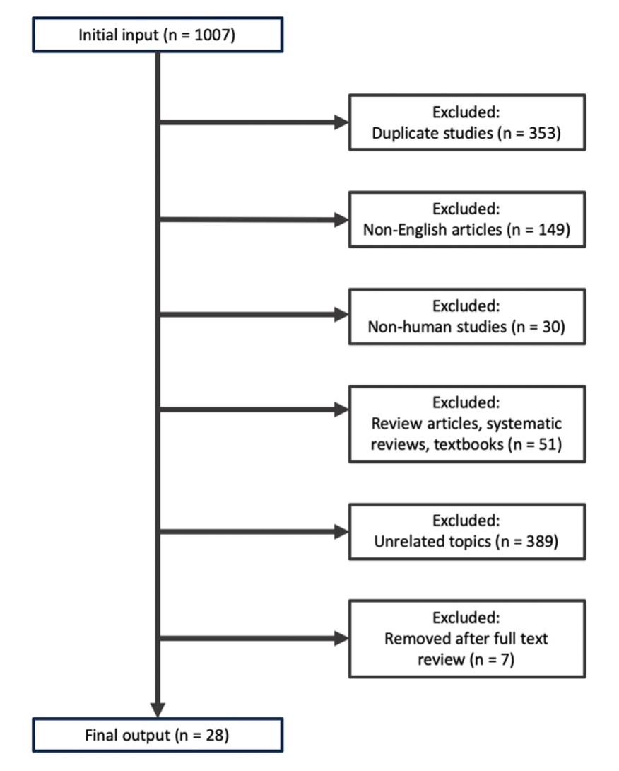
This systematic review was completed in 2020 using the Preferred Reporting Systems for Systematic Reviews and Meta-Analysis (PRISMA) guidelines.10 Preliminary searches were performed using data from PubMed, Embase, and Cochrane Library comprising studies dated through December 2019. The primary search included the keywords “tongue cancer”, “lingual cancer”, “buccal cancer”, and “metastasis”. A secondary search was done using the keywords “cardiac metastasis” and “tongue”.

The selected articles were combined to create a composite list of 1,007 studies to review. Studies that included review articles, textbooks, non-human subjects, non-English language, or unrelated topics were excluded. The inclusion criteria utilized in this systematic review are outlined in Figure 1.

Figure 1
Figure 1.Literature selection criteria. Literature selection methodology was constructed using the Preferred Reporting Items for Systematic Reviews and Meta-Analyses (PRISMA).

Study selection and data extraction

Each article within the composite list of studies was reviewed for inclusion by two independent authors (CK, TN). The titles and abstracts were screened for information regarding metastatic LSCC presentation, diagnosis, and management. Relevant articles were further examined via full text review and a finalized list was generated for in-depth analysis (Supplemental Table 1). A total of 31 cases of LSCC cardiac metastasis within 28 studies met inclusion criteria and were comprehensively reviewed (Supplemental Table 2).

All 28 selected studies had obtained an Oxford Centre for Evidence-Based Medicine (OCEBM) evidence categorization of Level 4 given their status as either a case report or case series. Relevant patient demographics, symptoms, LSCC history, clinical findings, treatment strategies, complications, and outcomes were collected. It was assumed death had occurred within six months if patients underwent palliative care.

RESULTS

Sample Demographics and Exposure

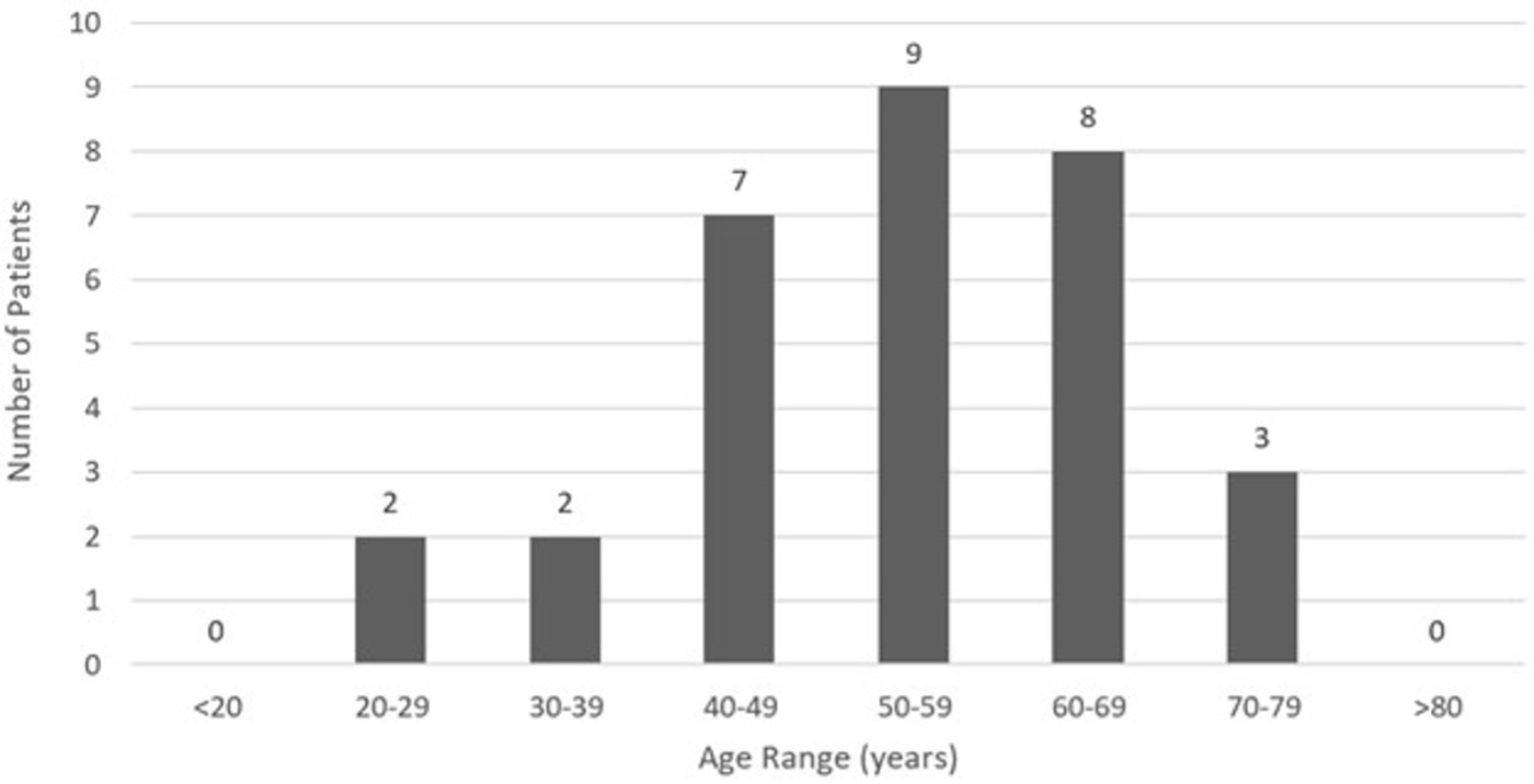
An analysis of demographic characteristics of patients in selected articles included 19 (61.3%) males and 12 (38.7%) females with a mean age of 53.6 (SD = 12.9) years ranging from 23.0 - 77.0 years. LSCC cardiac metastasis presented primarily between the ages of 40 - 69 (Figure 2). Average time from primary cancer diagnosis to cardiac metastasis identification was 2.2 (SD = 2.6) years, ranging from 0.2 - 11.0 years. Ten (32.3%) patients reported significant tobacco exposure and five (16.1%) patients admitted to alcohol use.

Figure 2
Figure 2.LSCC age distribution by decade of life.

Presenting Symptoms and Physical Examination

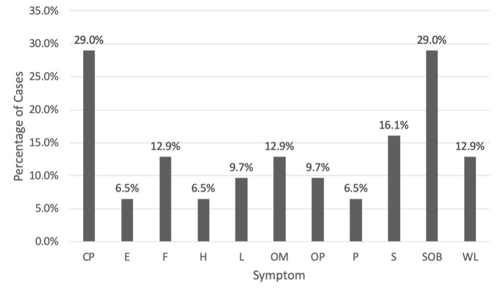
Chest pain and shortness of breath were the most common causes of initial presentation with nine (29.0%) cases, followed by five (16.1%) cases with syncope, four (12.9%) cases with weight loss, four (12.9%) cases with fever, four (12.9%) cases with oral mass, three (9.7%) cases with lymphadenopathy (i.e., enlargement of lymph nodes > or = 1), and three (9.7%) cases with oral pain. Other symptoms included two (6.5%) patients with edema, two (6.5%) patients with hemoptysis (i.e., blood mixed in sputum), and two (6.5%) patients with palpitations. A complete outline of patient symptomatology is depicted in Figure 3.

The most common physical examination findings included four (12.9%) patients with hypotension, four (12.9%) patients with fever, three (9.7%) patients with tachycardia, and three (9.7%) patients with cardiac murmur.

Figure 3
Figure 3:Symptomatology of LCCC Patients diagnosed with cardiac metastasis.

Abbreviations: CP = chest pain, E = edema, F = fever, H = hemoptysis, L = lymphadenopathy, OM = oral mass, OP = oral pain, P = palpitations, S = syncope, SOB = shortness of breath, WL = weight loss.

Cardiac Evaluation

Electrocardiogram (ECG) testing was reported in 19 (61.3%) patients, which found the most common abnormality to be ST-segment elevation in 12 (63.2%) patients (Supplemental Table 3). Other commonly reported ECG findings included arrhythmia in six (31.6%) patients, bundle branch block in four (21.1%) patients, t-wave inversion in four (21.1%) patients, and tachycardia in three (15.8%) patients. Troponin testing was reported in 31 patients with positive troponin elevations occurring in five (16.1%) cases.

Imaging Modalities and Findings

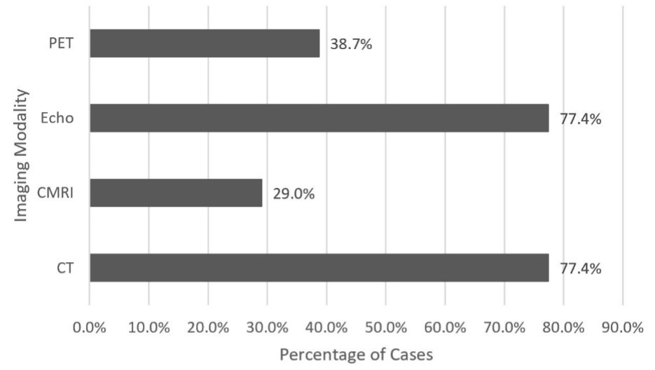
The most utilized imaging modalities were echocardiogram and computed tomography (CT) both occurring in 24 (77.4%) cases, followed by positron emission tomography (PET) in 12 (38.7%) cases, and cardiac magnetic resonance imaging (CMRI) in nine (29.0%) cases (Figure 4). On echocardiogram, the most common finding was pericardial effusion occurring in seven (29.2%) cases, followed by six (25.0%) cases with right ventricular outflow tract obstruction, three (12.5%) cases with valvular dysfunction, and two (8.3%) cases with wall motion abnormality (i.e., kinetic alterations in the cardiac wall motion taking place during the cardiac cycle) (Supplemental Table 3).

Of the patients with reported valvular dysfunction, two (66.6%) cases had impaired function of the pulmonic valve and one (33.3%) case had impaired tricuspid valve function. In the 12 patients who underwent PET scan, extra-cardiac uptake was most reported in the lung with five (41.7%) reported cases, followed by two (16.7%) cases in the bone, two (16.7%) cases in the liver, and one (8.3%) case in the muscle. The number of extra-cardiac foci ranged from 0.0 - 7.0 sites with an average of 1.8 (SD = 1.7) sites.

Figure 4
Figure 4:Utilization of diagnostic modalities. Four major imaging modalities were used to assist with the diagnosis of cardiac metastasis from LSCC.

Abbreviations: CMRI = cardiac magnetic resonance imaging; CT = computed tomography; Echo = echocardiogram; PET = positron emission tomography.

Location of Cardiac Metastasis

Out of 31 selected cases, 27 disclosed the number of cardiac metastases. Number of distinct cardiac metastases ranged from 1.0 - 4.0, with an average of 1.5 (SD = 0.8) metastases. Most cases presented with one individual mass in 17 (63.0%) cases, followed by two masses in 8 (29.6%) cases, and three or more masses in 2 (7.4%) cases.

The locations of metastases were distinctly specified in 30 of 31 cases. Right-sided metastases were most common with 23 (76.7%) instances, followed by 15 (50.0%) instances of left-sided metastases, and 11 (36.7%) instances of septal metastases. The most common location of metastasis was the right ventricle with 17 (56.7%) instances, followed by the left ventricle in 13 (43.3%) instances, interventricular septum in nine (30.0%) instances, and right atrium in six (20.0%) instances. This analysis also revealed eight (26.7%) patients with pericardial metastasis. Locational distinctions are further defined in Supplemental Table 2 and Supplemental Table 4.

Patient Management

Primary LSCC treatment strategies were discussed in 29 (93.5%) of 31 total cases. The most frequently reported primary treatment was surgery in 25 (86.2%) cases, followed closely by radiotherapy in 23 (79.3%) cases, and chemotherapy in 13 (44.8%) cases. The most common surgical intervention was neck dissection in eight (32.0%) cases, followed by partial glossectomy (i.e., removal of tongue tissue) in six (24.0%) cases, and hemi-mandibulectomy (i.e., removal of part of or all of the hemimandible, or one side or half of the mandible) in two (8.0%) cases.

Treatment strategies for metastatic lesions were described in 22 (71.0%) of 31 cases which included chemotherapy, surgical intervention, radiotherapy, immunotherapy, and palliative treatment. Given the poor prognosis, the most elected therapy was palliative treatment in 15 (68.2%) instances. Chemotherapy was the next most common intervention in nine (40.9%) instances, followed by surgery in four (18.2%) instances, radiotherapy in three (13.6%) instances, and immunotherapy in one (4.5%) instance.

Of those treated with chemotherapy, the most used agents were cisplatin in three (33.3%) cases and 5-fluorouracil in three (33.3%) cases. Other chemotherapeutics included bleomycin, etoposide, and methotrexate. Of note, immunotherapy with pembrolizumab was utilized in only one (4.5%) case. Individual treatments are further outlined in Supplemental Table 2.

Patient Outcomes

Twenty-two total cases (70.9%) reported outcome results, of which none showed remission of LSCC cardiac metastasis. Death was reported in 20 (90.9%) cases with two (9.1%) cases reporting palliative care and assumed death in less than six months. Of the 14 cases to report death within a known period, 9 (64.3%) reported death within one month of LSCC cardiac metastasis diagnosis.

Cause of death was listed in 16 cases which included eight (50.0%) deaths from cardiac metastasis, two (12.5%) deaths from sepsis, two (12.5%) deaths from arrhythmia/sudden cardiac death, one (6.3%) death from heart failure, one (6.3%) death from hemoptysis, one (6.3%) death from death in sleep, and one (6.3%) death from tumor embolism. Four (25.0%) cases had cardiac-related deaths, while 12 (75.0%) cases had tumor-specific or non-cardiac deaths.

DISCUSSION

Primary squamous cell carcinoma (SCC) accounts for more than 90% of cancer diagnoses within the head and neck region and is the sixth most common cancer overall by incidence.11 Classically, LSCC displays a predilection for the elderly male population,12 although it can present more aggressively in younger individuals with higher rates of metastasis and mortality.13 Recent research also suggests that young Caucasian females are demonstrating an overall increase in disease incidence.7 In this study, the majority of patients (51.6%) were males aged 40-69 years old, consistent with the typical presentation of LSCC.

In addition to age and sex, modifiable factors such as tobacco and alcohol exposure carry significant independent and synergistic risks for LSCC.14,15 Exposure to viral infections such as Epstein-Barr virus (EBV) and human papilloma virus (HPV) have also been associated with LSCC.16,17 In this systematic review, most cases failed to describe previous history of alcohol or tobacco use and no cases reported previous EBV or HPV exposure.

Cardiac metastasis of LSCC is typically diagnosed post-mortem on autopsy due to limited specific clinical manifestations.18 When diagnosed ante-mortem, patients may present with nonspecific cardiac symptoms such as chest pain, shortness of breath, and palpitations.19 In this systematic review, patients commonly demonstrated nonspecific cardiac symptoms such as chest pain, shortness of breath, syncope, and weight loss. Of these symptoms, chest pain and shortness of breath were the most prevalent (29.0% of cases), followed by syncope, then weight loss. Common physical exam findings were generally nonspecific (e.g., hypotension, fever, tachycardia, and cardiac murmur) in the selected articles.

Primary SCC originating from the head and neck with cardiac metastasis is extremely rare, as most cardiac neoplasms occur secondarily from areas such as the breast, lung, and esophagus.20 When present, cardiac metastasis most frequently occurs in the pericardium, followed by the myocardium, epicardium, endocardium, and intracavitary region.21

In this systematic review, right-sided cardiac metastasis was more common than left-sided metastasis, and ventricular involvement was more common than atrial involvement. The examined cases demonstrated a higher incidence of intracardiac tumors compared to pericardial tumors. Possible explanations for these findings relate to the hematogenous spread of squamous cell neoplasms through the coronary arteries, direct contiguous extension, and retrograde lymphatic flow.22

Since symptomatic presentations of LSCC cardiac metastasis can be highly variable, imaging studies are needed to assess the extent of cardiac involvement. First-line imaging for cardiac metastasis of LSCC begins with an echocardiogram due to its wide availability and lack of radiation.23 When there are no contraindications, contrast-enhanced CT scans are the diagnostic imaging modality of choice, as non-contrast CT scans may fail to identify small myocardial masses.24 If further imaging is indicated due to inconclusive CT results, CMRI is considered the most definitive imaging modality for evaluation of myocardial metastasis and delineation of intracardiac tumor thrombi.23

Cervical lymph node involvement is a strong prognostic factor when delineating patient outcomes for LSCC.25 Five-year survival rates for patients with oral SCC lymph node metastasis are low at 25-40%, which contrasts with a 90% survival rate for individuals without lymph node metastasis.12 Metastases to the heart further worsen this prognosis with a median survival of approximately 3.5 months without treatment.26–28 Palliative care is typically indicated for LSCC cardiac metastasis patients, as most cases present with poorly prognostic advanced cardiac SCC metastasis.26

During this review, no example of remission or significant survival was demonstrated in patients with cardiac metastasis of LSCC. Many cases resulted in cardiac-related death within 30 days of cardiac metastasis presentation and five deaths occurred upon initial admission for metastatic evaluation. These findings suggest that palliation may be the most appropriate treatment strategy for patients with LSCC cardiac metastasis given the poor prognosis with current treatment strategies.

Review Limitations

This systematic review had several limitations. First, only 31 cases of cardiac metastasis from LSCC met inclusion criteria which limits the power of this study. Second, LSCC cardiac metastasis may have been underreported in the medical literature due to missed diagnoses from lack of symptomatology or post-mortem follow-up. Third, the results of this study may not be completely representative of all documented cases of LSCC cardiac metastasis due to the omission of many non-English case reports and case series.

CONCLUSIONS

Cardiac metastasis from primary LSCC demonstrates a dangerous, uncommon presentation of malignancy. Clinical suspicion for LSCC cardiac metastasis should arise in patients with new onset chest pain and shortness of breath in the setting of prior diagnosis of LSCC. Prior tobacco and alcohol use should generate additional diagnostic speculation in these patients.

In the setting of previously known disease, advanced imaging such as echocardiogram, CT, and CMRI may prove useful for identification of disease progression. LSCC cardiac metastases typically favor the right and left ventricles, but are not exclusive to these sites. Due to the poor prognostic implications of LSCC cardiac metastasis, myocardial biopsy is unlikely to alter patient management and palliative discussions should be considered.


Conflict of Interest

None

Financial Support

None

Accepted: August 05, 2021 EDT

References

1.
Rodrigues VC, Moss SM, Tuomainen H. Oral cancer in the UK: To screen or not to screen. Oral Oncol. 1998;34(6):454-465. doi:10.1016/s1368-8375(98)00052-9
Google Scholar
2.
Sano D, Myers JN. Metastasis of squamous cell carcinoma of the oral tongue. Cancer Metastasis Rev. 2007;26(3-4):645-662. doi:10.1007/s10555-007-9082-y
Google Scholar
3.
Irani S. Distant metastasis from oral cancer: A review and molecular biologic aspects. J Int Soc Prev Community Dent. 2016;6(4):265-271. doi:10.4103/2231-0762.186805
Google ScholarPubMed CentralPubMed
4.
Jemal A, Clegg LX, Ward E, et al. Annual report to the nation on the status of cancer, 1975-2001, with a special feature regarding survival. Cancer. 2004;101(1):3-27. doi:10.1002/cncr.20288
Google Scholar
5.
Santos HB, dos Santos T, Paz A, et al. Clinical findings and risk factors to oral squamous cell carcinoma in young patients: A 12-year retrospective analysis. Med Oral Patol Oral y Cir Bucal. 2016;21(2):e151-156. doi:10.4317/medoral.20770
Google ScholarPubMed CentralPubMed
6.
Llewellyn CD, Johnson NW, Warnakulasuriya KA. Risk factors for squamous cell carcinoma of the oral cavity in young people — a comprehensive literature review. Oral Oncol. 2001;37(5):401-418. doi:10.1016/s1368-8375(00)00135-4
Google Scholar
7.
Ng JH, Iyer NG, Tan MH, Edgren G. Changing epidemiology of oral squamous cell carcinoma of the tongue: A global study. Head Neck. 2017;39(2):297-304. doi:10.1002/hed.24589
Google Scholar
8.
Pattni N, Rennie A, Hall T, Norman A. Cardiac metastasis of oral squamous cell carcinoma. BMJ Case Rep. 2015;2015:bcr2015211275. doi:10.1136/bcr-2015-211275
Google ScholarPubMed CentralPubMed
9.
Kim JK, Sindhu K, Bakst RL. Cardiac metastasis in a patient with head and neck cancer: A case report and review of the literature. Case Rep Otolaryngol. 2019;2019:1-8. doi:10.1155/2019/9581259
Google ScholarPubMed CentralPubMed
10.
Moher D, Liberati A, Tetzlaff J, Altman DG, for the PRISMA Group. Preferred reporting items for systematic reviews and meta-analyses: The PRISMA statement. BMJ. 2009;339:b2535. doi:10.1136/bmj.b2535
Google ScholarPubMed CentralPubMed
11.
Mendelsohn AC, Gray JE, Howley A, et al. The Molecular Basis of Cancer. Saunders; 2015.
Google Scholar
12.
Noguti J, De Moura CF, De Jesus GP, et al. Metastasis from oral cancer: An overview. Cancer Genomics and Proteomics. 2012;9(5):329-336.
Google Scholar
13.
Jeon JH, Kim MG, Park JY, et al. Analysis of the outcome of young age tongue squamous cell carcinoma. Maxillofac Plast Reconstr Surg. 2017;39(1):41. doi:10.1186/s40902-017-0139-8
Google ScholarPubMed CentralPubMed
14.
Bachar G, Hod R, Goldstein DP, et al. Outcome of oral tongue squamous cell carcinoma in patients with and without known risk factors. Oral Oncol. 2011;47(1):45-50. doi:10.1016/j.oraloncology.2010.11.003
Google Scholar
15.
Subapriya R, Thangavelu A, Mathavan B, Ramachandran CR, Nagini S. Assessment of risk factors for oral squamous cell carcinoma in Chidambaram, southern India: A case-control study. Eur J Cancer Prev. 2007;16(3):251-256. doi:10.1097/01.cej.0000228402.53106.9e
Google Scholar
16.
Guidry JT, Birdwell CE, Scott RS. Epstein-Barr virus in the pathogenesis of oral cancers. Oral Dis. 2018;24(4):497-508. doi:10.1111/odi.12656
Google ScholarPubMed CentralPubMed
17.
Ragin CC, Modugno F, Gollin SM. The epidemiology and risk factors of head and neck cancer: A focus on human papillomavirus. J Dent Res. 2007;86(2):104-114. doi:10.1177/154405910708600202
Google Scholar
18.
Delabie P, Evrard D, Zouhry I, et al. Squamous cell carcinoma of the tongue with cardiac metastasis on 18F-FDG PET/CT: A case report and literature review. Medicine (Baltimore). 2021;100(15):e25529. doi:10.1097/md.0000000000025529
Google ScholarPubMed CentralPubMed
19.
Malekzadeh S, Platon A, Poletti PA. Cardiac metastasis of tongue squamous cell carcinoma complicated by pulmonary embolism. Medicine (Baltimore). 2017;96(28):e7462. doi:10.1097/md.0000000000007462
Google ScholarPubMed CentralPubMed
20.
Tandon V, Kethireddy N, Balakumaran K, Kim AS. Metastatic squamous cell carcinoma to the heart: An unusual cause of ST elevation—a case report. Eur Heart J Case Rep. 2019;3(2):ytz029. doi:10.1093/ehjcr/ytz029
Google ScholarPubMed CentralPubMed
21.
Scott RW, Garvin CF. Tumors of the heart and pericardium. Am Heart J. 1939;17(4):431-436. doi:10.1016/s0002-8703(39)90593-4
Google Scholar
22.
Werbel GB, Skom JH, Mehlman D, Michaelis LL. Metastatic squamous cell carcinoma to the heart. Unusual cause of angina decubitus and cardiac murmur. Chest. 1985;88(3):468-469. doi:10.1378/chest.88.3.468
Google Scholar
23.
Lichtenberger JP, Reynolds DA, Keung J, Keung E, Carter BW. Metastasis to the heart: A radiologic approach to diagnosis with pathologic correlation. Am J Roentgenol. 2016;207(4):764-772. doi:10.2214/ajr.16.16148
Google Scholar
24.
Zitzelsberger T, Eigentler TK, Krumm P, et al. Imaging characteristics of cardiac metastases in patients with malignant melanoma. Cancer Imaging. 2017;17(1):19. doi:10.1186/s40644-017-0122-8
Google ScholarPubMed CentralPubMed
25.
Leemans CR, Tiwari R, Nauta J, Van der Waal I, Snow GB. Regional lymph node involvement and its significance in the development of distant metastases in head and neck carcinoma. Cancer. 1993;71(2):452-456. doi:10.1002/1097-0142(19930115)71:2
Google Scholar
26.
Reynen K, Köckeritz U, Strasser RH. Metastases to the heart. Ann Oncol. 2004;15(3):375-381. doi:10.1093/annonc/mdh086
Google Scholar
27.
Takenaka S, Hashimoto N, Araki N, et al. Eleven cases of cardiac metastases from soft-tissue sarcomas. Jpn J Clin Oncol. 2011;41(4):514-518. doi:10.1093/jjco/hyq246
Google Scholar
28.
Fotouhi Ghiam A, Dawson LA, Abuzeid W, et al. Role of palliative radiotherapy in the management of mural cardiac metastases: Who, when and how to treat? A case series of 10 patients. Cancer Med. 2016;5(6):989-996. doi:10.1002/cam4.619
Google ScholarPubMed CentralPubMed
29.
Nagata S, Ota K, Nagata M, Shinohara M. Cardiac metastasis of head and neck squamous cell carcinoma. Int J Oral Maxillofac Surg. 2012;41(12):1458-1462. doi:10.1016/j.ijom.2012.07.017
Google Scholar
30.
Onwuchekwa J, Banchs J. Early cardiac metastasis from squamous cell carcinoma of the tongue in 2 patients. Tex Heart Inst J. 2012;39(4):565-567.
Google Scholar
31.
Browning CM, Craft JF, Renker M, Joseph Schoepf U, Baumann S. Squamous cell carcinoma of the tongue with metastasis to the right ventricle. Am J Med Sci. 2015;349(5):461-462. doi:10.1097/maj.0000000000000411
Google Scholar
32.
McKeag N, Hall V, Johnston N, et al. Cardiac metastasis from a squamous cell carcinoma of the tongue in the absence of local recurrence. Ulster Med J. 2013;82(3):193-194.
Google Scholar
33.
Hans S, Chauvet D, Sadoughi B, Brasnu DF. Cardiac metastasis after squamous cell carcinoma of the base of tongue. Am J Otolaryngol. 2009;30(3):206-208. doi:10.1016/j.amjoto.2008.03.008
Google Scholar
34.
Kumar D, Mankame P, Sabnis G, Nabar A. A case report: metastatic complete heart block. Eur Heart J Case Rep. 2018;2(4):yty131. doi:10.1093/ehjcr/yty131
Google ScholarPubMed CentralPubMed
35.
Nanda A, Khouzam RN, Jefferies J, Moon M, Makan M. Right heart mass in transit with a hemorrhagic pericardial effusion: A diagnostic dilemma. Cureus. 2019;11(2):e4009. doi:10.7759/cureus.4009
Google ScholarPubMed CentralPubMed
36.
Werbel GB, Skom JH, Mehlman D, Michaelis LL. Metastatic squamous cell carcinoma to the heart. Chest. 1985;88(3):468-469. doi:10.1378/chest.88.3.468
Google Scholar
37.
Puranik AD, Purandare NC, Sawant S, et al. Asymptomatic myocardial metastasis from cancers of upper aero-digestive tract detected on FDG PET/CT: A series of 4 cases. Cancer Imaging. 2014;14(1):16. doi:10.1186/1470-7330-14-16
Google ScholarPubMed CentralPubMed
38.
Makhija Z, Deshpande R, Desai J. Unusual tumours of the heart: Diagnostic and prognostic implications. J Cardiothorac Surg. 2009;4(1). doi:10.1186/1749-8090-4-4
Google ScholarPubMed CentralPubMed
39.
Wadler S, Muller R, Spigelman MK, Biller H. Fulminant disseminated carcinomatosis arising from squamous cell carcinoma of the tongue. Am J Med. 1985;78(1):149-152. doi:10.1016/0002-9343(85)90476-0
Google Scholar
40.
Shafiq A, Samad F, Roberts E, Tajik AJ. Squamous cell carcinoma of the tongue with metastasis to the myocardium: A rare occurrence. J Am Coll Cardiol. 2019;73(9):2258. doi:10.1016/s0735-1097(19)32864-5
Google Scholar
41.
Dredla B, Siegel J, Jaeckle K. BM-12: Cerebral infarction secondary to pulmonary vein compression and left atrial appendage tumor infiltration as the presenting sign of metastatic squamous cell carcinoma of the base of the tongue. NeuroOncol. 2014;16(suppl 5):v34. doi:10.1093/neuonc/nou240.12
Google ScholarPubMed Central
42.
Yadav NU, Gupta D, Baum MS, Roistacher N, Steingart RM. Cardiac metastases from head and neck cancer mimicking a myocardial infarction. J Oral Maxillofac Surg. 2014;72(8):1627-1635. doi:10.1016/j.joms.2014.02.015
Google Scholar
43.
Zatuchni J, Burris A, Vejviboonsom P, Voci G. Metastatic epidermoid cardiac tumor manifested by persistent ST segment elevation. Am Heart J. 1981;101(5):674-675. doi:10.1016/0002-8703(81)90238-6
Google Scholar
44.
Shafiq A, Samad F, Roberts E, Levin J, Nawaz U, Tajik AJ. Squamous cell carcinoma of the tongue with metastasis to myocardium: Report of a case and literature review. Case Rep Cardiol. 2019;2019:1-6. doi:10.1155/2019/1649580
Google ScholarPubMed CentralPubMed
45.
Kim JK, Sindhu K, Bakst RL. Cardiac metastasis in a patient with head and neck cancer: A case report and review of the literature. Case Rep Otolaryngol. 2019;2019:1-8. doi:10.1155/2019/9581259
Google ScholarPubMed CentralPubMed
46.
Demir V, Ede H, Hidayet S, Turan Y. Right ventricular metastasis producing electrocardiographic changes mimicking acute ST elevation myocardial infarction: a case of misdiagnosis. Am J Cardiol. 2018;121(8):e131. doi:10.1016/j.amjcard.2018.03.299
Google Scholar
47.
Chua S, Liu WH, Lee WC. Isolated huge right ventricular tumor: Cardiac metastasis of tongue cancer. Korean J Intern Med. 2017;32(6):1119-1120. doi:10.3904/kjim.2016.143
Google ScholarPubMed CentralPubMed
48.
Duband S, Paysant F, Scolan V, Forest F, Péoc’h M. Sudden death due to myocardial metastasis of lingual squamous cell carcinoma. Cardiovasc Pathol. 2011;20(4):242-243. doi:10.1016/j.carpath.2010.07.002
Google Scholar
49.
Ito T, Ishikawa N, Negishi T, Ohno K. Cardiac metastasis of tongue cancer may cause sudden death. Auris Nasus Larynx. 2008;35(3):423-425. doi:10.1016/j.anl.2007.04.017
Google Scholar
50.
Rivkin A, Meara JG, Li KK, Potter C, Wenokur R. Squamous cell metastasis from the tongue to the myocardium presenting as pericardial effusion. Otolaryngol Head Neck Surg. 1999;120(4):593-595. doi:10.1053/hn.1999.v120.a84489
Google Scholar
51.
Matsuyama M, Ito M, Baba S. Lingual cancer with myocardial metastasis. Ann Otol Rhinol Laryngol. 1963;72(1):56-61. doi:10.1177/000348946307200105
Google Scholar
52.
Cabot RC, Castleman B, McNeely BU, Kranes A, Scully RE. Case records of the Massachusetts General Hospital. Weekly clinicopathological exercises. Case 8-1971. N Engl J Med. 1971;284(8):435-442. doi:10.1056/nejm197102252840809
Google Scholar
53.
Reddy G, Ahmed MI, Lloyd SG, Brott BC, Bittner V. Left anterior descending coronary artery occlusion secondary to metastatic squamous cell carcinoma presenting as ST-segment-elevation myocardial infarction. Circulation. 2014;129(24):e652-653. doi:10.1161/circulationaha.114.009157
Google Scholar
54.
Ewald FW, Scherff AH. A 60-year-old man with a malignant tumor of the upper airway and unexplained respiratory failure. Chest. 1997;111(1):239-241. doi:10.1378/chest.111.1.239
Google Scholar

Supplementary Tables

Supplemental Table 1.Level of evidence and conclusion of studies.
Authors Study Year LOE (1a-5) Study Design Subjects Authors’ Conclusions
Tandon20 2019 4 CR 1 Patients with current malignancies in addition to cardiac symptoms and/or ECG changes should warrant investigation for possible cardiac metastasis. Different imaging modalities such as CMRI, PET-CT, and echocardiography play an important role in diagnosis and management.
Nagata29 2012 4 CR 1 Cardiac metastasis from LSCC is typically a difficult diagnosis due to the lack of clinical findings in the early stages. However, as the metastases become more advanced, clinical signs may occur, resulting in a variety of cardiac pathologies. Many of the cardiac sequelae result in ECG changes, emphasizing the importance of ECG testing in these patients.
Onwuchekwa30 2012 4 CS 2 These cases show the importance of evaluating the heart with echocardiography to assess for cardiac metastasis in patients with head and neck cancer and confirmed metastatic disease.
Browning31 2015 4 CR 1 It may be necessary to evaluate patients with lingual malignancies with imaging to rule out metastases. Lingual squamous cell carcinoma metastases are commonly asymptomatic, and thus, imaging is important in establishing the diagnosis.
McKeag32 2013 4 CR 1 This case shows that cardiac metastasis from LSCC can present without symptoms or characteristic metastasis examination findings such as lymphadenopathy.
Hans33 2009 4 CR 1 Clinicians may consider the potential for cardiac metastases in patients with a history of head and neck malignancies who display new-onset cardiovascular symptoms. These patients typically have a very poor prognosis.
Kumar34 2018 4 CR 1 Clinicians should consider cardiac metastasis and hypercalcemia-induced rhythm disturbances in patients with a history of LSCC malignancy or current LSCC malignancy and concurrent ECG changes. It may be necessary to check calcium levels and treat the patient if levels are abnormal.
Nanda35 2019 4 CR 1 This case shows the importance of an initial echocardiogram in addition to the integration of PET and CT scans to detect LSCC cardiac metastasis.
Werbel36 1985 4 CR 1 This case demonstrates the importance of using echocardiography in patients with LSCC and atypical angina and/or a new heart murmur. Echocardiography is a good initial step to look for cardiac metastases.
Puranik37 2014 4 CS 2 In asymptomatic patients with a history of cancer, whole body dual imaging with PET-CT can show undiagnosed metastatic sites.
Makhija38 2009 4 CR 1 This case report stresses the lack of appropriate screening protocols for patients with LSCC and possible cardiac metastases. The report recommends a transesophageal echocardiogram as the best initial screen, as it is a sensitive test.
Wadler39 1985 4 CR 1 This case report emphasizes that there are many other factors besides morphology and anatomic extent that can be used to determine LSCC prognosis.
Shafiq40 2019 4 CR 1 This case report shows that the lack of symptoms in cardiac metastasis from LSCC can result in delayed cardiac imaging and subsequently delayed diagnosis.
Dredla41 2014 4 CR 1 Patients with a history of cancer who have a cerebrovascular accident should have cardiac imaging to rule out a cardiac cause, such as cardiac metastases.
Yadav42 2014 4 CR 1 This case highlights the importance of an ECG evaluation in patients with a cancer diagnosis. This can provide diagnostic clues for potential cardiac metastases. Diagnostic confirmation can be done using multiple imaging modalities, such as echocardiography, CT scan, and CMRI.
Zatuchni43 1981 4 CS 2 LSCC cardiac metastasis can result in myocardial or pericardial injury, shown by ST-elevations on ECG. ST-elevation may be a potential sign of cardiac metastasis.
Shafiq44 2019 4 CR 1 This case emphasizes the importance of the clinician maintaining a high index of suspicion to identify patients with metastatic LSCC. A high risk patient may need surveillance imaging with echocardiography, CMRI, and/or PET-CT.
Kim45 2019 4 CR 1 LSCC patients with cardiac symptoms may benefit from a multimodal approach consisting of PET-CT, CMRI, echocardiography, and ECG. These imaging modalities are used to confirm the diagnosis and establish the location and extent of disease, which can guide management.
Demir46 2018 4 CR 1 This case shows the importance of keeping myocardial metastases on the differential diagnosis when patients with known malignancies present with ECG changes. Cardiac metastases can sometimes present like an acute myocardial infarction on ECG testing.
Chua47 2017 4 CR 1 LSCC is more common in Asian countries. It is always important to consider metastasis of LSCC to the heart and other organs, especially if patients with known LSCC present with cardiac symptoms.
Duband48 2011 4 CR 1 Clinicians may consider an initial cardiac evaluation and serial follow-up ECGs in patients with SCC of the base of the tongue.
Ito49 2007 4 CR 1 Sudden death in patients with LSCC should prompt clinicians to consider the potential of cardiac metastases.
Rivkin50 1999 4 CR 1 Patients with cancer who develop new cardiac symptoms should prompt the clinician to evaluate for potential cardiac metastases. Evaluation should consist of echocardiography and/or CMRI. Treatments are typically only palliative, as cardiac metastases possess a poor prognosis.
Matsuyama51 1963 4 CR 1 This case reports a rare finding of widespread LSCC metastasis with cardiac metastases isolated to the myocardium. This is a rare finding since most metastases affect the pericardium.
Malekzadeh19 2017 4 CR 1 Patients with a history of head and neck malignancy who present with cardiac symptoms may prompt the clinician to evaluate for cardiac metastases.
Cabot52 1971 4 CR 1 There are often no clinical signs of primary LSCC or metastatic LSCC to the heart, making the ante-mortem diagnosis very difficult.
Reddy53 2014 4 CR 1 Management of tumor-induced acute coronary occlusion is similar to acute coronary syndrome occlusion, utilizing coronary angiography and percutaneous coronary intervention.
Ewald54 1997 4 CR 1 Patients with a history of malignancy and unexplained arrhythmias or ECG changes may prompt the clinician to consider cardiac metastases on the differential diagnosis. Cardiac metastasis may present similarly to many types of cardiac pathology.

Abbreviations: CMRI = cardiac magnetic resonance imaging; CR = case report; CS = case series; CT = computed tomography; ECG = electrocardiogram; LSCC = lingual squamous cell carcinoma; PET = positron emission tomography; PET-CT = positron emission tomography-computed tomography; SCC = squamous cell carcinoma.

Supplemental Table 2.Detailed Patient Information.
Age/Sex Primary Location Signs & Symptoms Metastasis Locations First Identifying Imaging Primary Treatment Metastasis Treatment Outcomes
M 59 Right tongue mass Fever LA & pericardium CT & Echo Partial glossectomy, right RND, CTX Cardiac surgery Death 3 weeks after surgery
F 45 Tongue Syncope & dyspnea RV, LV, IVS CTA & Echo Partial right glossectomy, RND, RT. RT (brain) NR
F 36 Tongue Palpitations & SOB LV CT & Echo CTX-RT, partial left glossectomy, and left neck LN dissection RT Death 2 months after
M 50 Base of tongue Odynophagia, otalgia, dysphonia, WL, tongue and neck pain Apex of RV PET-CT 2 months radiation, total glossectomy & bilateral RND None NR
M 77 NR Recurrent syncope RA & RV infiltration of myocardium Echo None None Death 28 days after diagnosis
M 54 Tongue base Dyspnea, LE edema, & hemoptysis RV CT & Echo Glossectomy, left RND, cisplatin, 5-FU None Death
F 23 Tongue CP, Right arm paresthesias, cough, SOB LV & RV infiltration of myocardium Echo Surgical resection with clear margins None NR
M 28 Tongue Recurrent syncope RA, LV, IVS Echo Hemimandibulectomy & post-operative RT Pacemaker implantation Death 5 days after pacemaker placement
M 47 Tongue Dizziness, SOB, chest tightness, night sweats RA, RV, pulmonary artery CT chest Surgical excision with CTX-RT None Patient lost to follow-up
F 61 Base of tongue CP, palpitations RA & pericardium Echo Hemiglossectomy with excisional biopsy of digastric LN. Neck RT None Death 7 weeks after cardiac diagnosis
F 32 Right. lateral tongue Nasal swelling LV & IVS PET-CT scan Wide excision & right lateral neck dissection CTX Follow-up CT stable
M 46 Right vallecula N/A (Follow-up) RV Endoscopy CTX-RT CTX Patient lost to follow-up
F 66 Tongue Anginal symptoms RV Coronary angiogram Radical resection None NR
M 57 Anterior tongue Fever, dyspnea Epicardium, myocardium, endocardium Echo Partial glossectomy CTX Death 14 days after admission from sepsis
M 43 Tongue N/A (Follow-up) Apex of LV & RV CT & Echo CTX-RT CTX Death due to cardiac metastasis
M 61 Left tongue base Cerebral infarction symptoms LAA and pulmonary vein CT & Echo CTX-RT None NR
M 76 Tongue Pneumonia RV, LV CT chest Partial glossectomy, multiple neck dissection, CTX Palliative care Died in one month
M 61 Tongue CP, dyspnea IVS and pericardium 99mTC scan RAD None Death
M 62 Tongue Malaise Diffuse myocardium 99mTC scan NS None Death
M 43 Right tongue N/A (Follow-up) RV, LV CT chest Right neck dissection & tongue resection, free flap reconstruction, CTX-RT CTX-RT Death
F 46 Left lateral tongue Left arm, ear, and throat pain LV, pericardium CT Hemiglossectomy, bilateral neck dissection, CTX-RT CTX-RT NR
M 59 Tongue Chest discomfort RV Echo Resection, RT None Death
M 63 Tongue Dyspnea RV Echo Resection and reconstruction None NR
F 57 Tongue Death RA, AV sulcus Autopsy Hemiglossectomy, bilateral cervical lymphadenectomy, CTX-RT None Death
M 66 Right tongue into oropharynx Death LV, IVS Autopsy Total glossolaryngectomy and dissection LN bilaterally None Death
M 57 Right tongue base Lower extremity edema RV Echo, ECG, CMRI, CT Local excision with post-operative RT to lesion and neck CTX Death
F 48 Anterior tongue CP, dyspnea, hemoptysis, anorexia. Diffuse myocardium, RA, LV, aortic valve NR Anterior tongue glossectomy, excision of submandibular LN None Death
F 58 Tongue CP NR CT Hemiglossectomy and RT Palliative CTX and immunotherapy Death
F 70 Tongue Fever & chills Diffuse pericardium CT Left hemiglossectomy, RND Thoracotomy Death
F 52 Tongue Dull CP RV, LV, IVS PET Hemiglossectomy and RND Pericardial window and palliative care Life expectancy < 3 months
M 60 Base of tongue Malnutrition, pneumonia Anterior wall of RV & pulmonic valve Echo RT and a composite tongue-jaw-neck resection Palliative care Death

Abbreviations: 5-FU = 5-fluorouracil; 99mTC = Technetium 99; CMRI = cardiac magnetic resonance imaging; CP = chest pain; CT = computed tomography; CTA = computed tomography angiography; CTX = chemotherapy; CTX-RT = chemotherapy/radiation; Echo = echocardiogram; IVS = interventricular septum; LA = left atrium; LAA = left atrial appendage; LE = lower extremity; LN = lymph node; LV = left ventricle; MRI = magnetic resonance imaging; NR = not recorded; NS = non-surgical; PET = positron emission tomography; PET-CT = positron emission tomography-computed tomography; RA = right atrium; RND = radical neck dissection; RT = radiation therapy; RV = right ventricle; SOB = shortness of breath; WL = weight loss.

Supplemental Table 3.Clinical patient characteristics.
Characteristics Data
No. of subjects 31
Male, n (%) 19 (61.3)
Female, n (%) 12 (38.7)
Mean age of diagnosis, n years (range) 53.6 (23.0-77.0)
Mean primary to metastasis diagnosis, n years (range) 2.2 (0.2-11.0)
Presenting symptoms, n (%)
Chest pain 9 (29.0)
Shortness of breath 9 (29.0)
Syncope 5 (16.1)
Weight loss 4 (12.9)
Fever 4 (12.9)
Oral mass 4 (12.9)
Lymphadenopathy 3 (9.7)
Oral pain 3 (9.7)
Edema 2 (6.5)
Hemoptysis 2 (6.5)
Palpitations 2 (6.5)
Physical exam findings, n (%)
Hypotension 4 (12.9)
Fever 4 (12.9)
Cardiac murmur 3 (9.7)
Tachycardia 3 (9.7)
Diagnostic findings, n (%)
Electrocardiography 19 (61.3)
ST elevation 12 (63.2)
Arrhythmia 6 (31.6)
Bundle branch block 4 (21.1)
T-wave inversion 4 (21.1)
Echocardiography 24 (77.4)
Pericardial effusion 7 (29.2)
Right ventricular outflow tract obstruction 6 (25.0)
Valvular dysfunction 3 (12.5)
Wall motion abnormality 2 (8.3)
Supplemental Table 4.Tumor location.
Characteristics Data
Primary tumor site, n (%)
Tongue 31 (100.0)
Cardiac metastases, n (%) (1 case omitted) 30 (96.8)
Right-sided metastasis 23 (76.7)
Left-sided metastasis 15 (50.0)
Septal metastasis 11 (36.7)
Pericardial metastasis 8 (26.7)
Specific location
Right ventricle 17 (56.7)
Left ventricle 13 (43.3)
Interventricular septum 9 (30.0)
Right atrium 6 (20.0)
Left atrium 2 (6.7)